Wednesday, December 14, 2016

Tamponade and Tautologies

Note:
Our case report Low-Pressure Pericardial Tamponade: Case Report and Review of the Literature was just published. I can't really plagarize that text, or use the images from the case, but I had the "good fortune" to see a second case of what I believe was low-pressure tamponade. So, I get to write a #FOAMed version, as well as a conventional publication! Wins all around.


“Tautology: a statement that is true by virtue of its logical form.”

 Or, as put by XKCD:
Tamponade, oddly, can be harder to define than tautology!

Of course, many people say things like “tamponade is defined clinically,” or cite a cut-off level of pulsus paradoxus. But such criteria can be either vague, arbitrary, or poorly supported by good evidence. (For a good review of myriad such issues, check out Misconceptions and facts about pericardial effusion and tamponade. Paywall – sorry.)

A lack of IVC plethora rules out tamponade... right?
Instead, focus (FoCUS?) on a simpler element of defining tamponade – the IVC. A large number of textbooks and reviews have stated that plethora of the IVC is a very sensitive feature of tamponade. For example, a beautiful recent handbook of focused ultrasound states that:

“If tamponade is a consideration, it may effectively be ruled out by demonstrating of 50% or more with deep inspiration without having to pursue more sophisticated echocardiographic techniques.”

A similarly strong statement about the role of the IVC was delivered by Ultrasound Podcast: Pericardial Tamponade:

“If the IVC is collapsing, you are almost certainly not dealing with tamponade.”

Let’s see a case, though, to point out the complexities behind such blanket statements.

Case: Recurrent pericardial effusion s/p pericardial window.

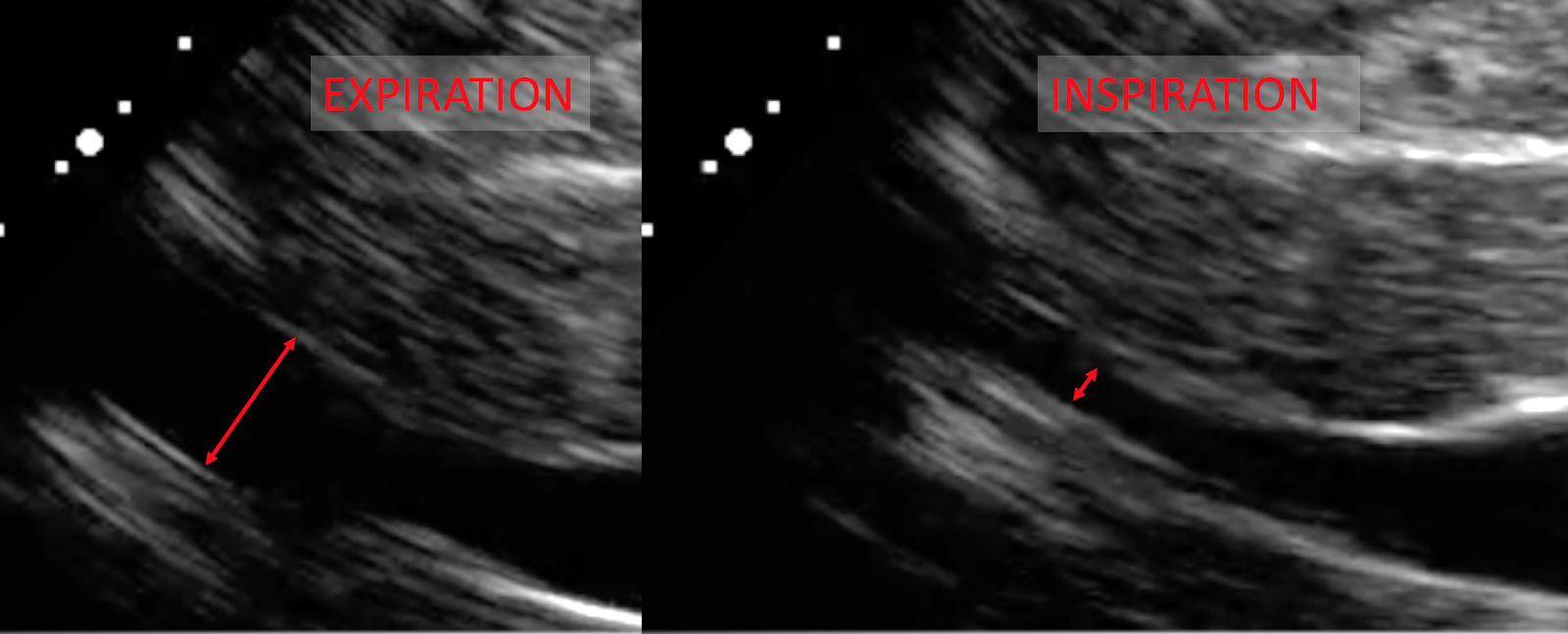
A patient presented to the ED with positional chest pain. However, she also had a recent history of pericardial effusion subsequent to a CABG. Although she had undergone a pericardial window at that time, and although her vital signs (aside from a modest tachypnea) were normal, the ED physician performed an echo. First, the sagittal and transverse views of the IVC:

Sagital
Transverse

And the clip:

The transverse images show that the IVC diameter is just < 2.1 cm, and collapses just about 50%. The sagittal images, however, demonstrate a much higher degree of collapse in the proximal IVC. Based on these views, could we have ruled-out tamponade?

Well, here are the rest of the views of the heart.
Screen Shot 2016-12-13 at 2.29.03 PM.png


  • Significant  effusion – check.   
  • RV collapse in diastole – check.
  • RA collapse in systole – check
  • Large respiratory variation in the MV inflow – check!

So, is this tamponade? Heck, yes! The next day she had 700 ml worth of effusion aspirated, with an initial intrapericardial pressure of 24 mm Hg. Pretty diagnostic.

But isn’t IVC plethora supposed to be almost 100% sensitive for tamponade?
Technically, 97% sensitive. Almost every textbook or review that gives this figure cites the same 1988 paper by Himelman et al., and it is imperative to understand the methods of that study before repeating that “almost 100% sensitive” claim.

Himelman et al. defined IVC plethora as < 50% IVC collapse during a deep inspiration, measured 2 cm distal to the RA entrance. So, plethora was not seen in our patient - was this a “miss” for detecting tamponade?

Well, Himelman had defined tamponade in a pretty narrow sense. They required that either:
  • a) The diastolic filling pressures (i.e. the diastolic pressure in the RA, RV, etc.) had to all be > 12 mm Hg), and be within 5 mm Hg of each other;
or
  • b) Hypotension was present, with an initial SBP < 100 mm Hg, increasing by ≥ 20 mm Hg immediately after aspiration.
Given this definition, our patient did not have tamponade during the ED evaluation!  **

  • First off, the SBP was never below 100 mm Hg.
  • Second, the bedside echo showed an IVC diameter < 2.1 cm, with > 50% inspiratory collapse, suggesting a RA pressure of only 0 - 5 mm Hg (per ASE guidelines),
There is a circular element to the inclusion criteria and the outcomes here...

The tautologic definition of tamponade is tautological.
Let’s dwell on Himelman’s definition of tamponade a bit more, and implications for the results they found.

They only included patients with either overt hypotension from their effusion, or patients with RAP ≥ 12 mm Hg. Of the 33 patients found to have tamponade, 20 had a RAP > 12 mm Hg. The ASE guidelines above, then, suggest that most of these patients would be expected to have an IVC that was fat and plethoric. And vice-versa: patients with a plethoric IVC would be predicted to have RA and intrapericardial pressures > 10, and likely > 12 for many of them.

Tautology: If we define tamponade as RA pressure > 12 mm Hg, any sign that defines a high RA pressure (IVC collapse < 50%) will be very common, by definition! 

Put another way, if you say that (based on Himelman) a non-plethoric IVC rules out tamponade, but only if you define tamponade such that the IVC should be plethoric.

But isn’t that a common-sense part of defining tamponade; JVD, pulsus, and hypotension? Well, it turns out that tamponade can be defined in many different ways.

There is more to tamponade than the end-stage, “clinical tamponade” presentation.
Studies of pericardial tamponade have used a variety of definitions of tamponade. And when different definitions of tamponade are used, the utility of IVC plethora (or JVD, which we can use as a clinical surrogate for IVC plethora) will vary as well.  For example:

  • A 1995 study defined tamponade as an “effusion causing dyspnea which is relieved by aspiration of the effusion,” and found that 87% of patients who received pericardiocentesis had significant relief of dyspnea. But half of the patients who fulfilled their definition of tamponade had no JVD (they did not evaluate IVC plethora).
  • A 2006 study looked at patients with pericardial effusions, all of whom received both right-heart catheterization and pericardiocentesis. Tamponade was defined solely as equalization of RA and IP pressures. They found that 24% of the cases of tamponade showed fairly low RA and IP pressures (< 7 mm Hg) prior to pericardiocentesis. Accordingly, only a fifth of these “low-pressure” cases of tamponade presented with JVD. This suggests that IVC plethora would have been correspondingly low as well, but echo results were not included, sadly.
  • A 2012 chart review also used a broader definition of tamponade than had Himelman (any of following echo features: characteristic RA, RV, or LV collapse; characteristic variation in blood flow through the TV or MV, or dilated IVC with lack of inspiratory collapse). Only 13% of these patients had a dilated, plethoric IVC.

Well, those studies didn’t look at “real” tamponade.
Because of studies like those just summarized, a subset of tamponade, know as “low pressure” tamponade, has been more widely recognized. Often seen when a patient with a pericardial effusion is also hypovolemic (e.g. from diuretics or illness), a low-pressure tamponade nonetheless manifests with the symptoms and hemodynamic characteristics of “real” tamponade. JVD, pulsus, or overt hypotension, however, may be initially absent.
Screen Shot 2016-12-14 at 11.33.50 AM.png
In fact, many experts have tried to push the view of tamponade as a spectrum, with broader inclusion criteria. Instead of a binary diagnosis of tamponade, we ought, they argue, to be evaluating where the patient is on the spectrum, as well as their potential to move “up” that spectrum; i.e deteriorate.

The IVC is but a single data point
In our patient, a modest degree of tachypnea, as well as a classic echo profile suggested that this patient was in echocardiographic tamponade, and at risk of deteriorating. If the non-plethoric IVC had been used to classify our patient simply as “not real tamponade,” the patient could have received less scrutiny, delayed evaluation, and perhaps a poorer outcome.

But this is nothing surprising, really. Almost every disease comes on a spectrum, and tamponade is no different. Accordingly, it isn’t useful, and may indeed be harmful, to teach that the IVC can “rule out tamponade.”
__________________________________________
** Yes, the intrapericardial/RA pressure was higher the next day, when directly measured during aspiration. However, the procedure was about 1 day after the echo. In that  interval probably more fluid had accumulated in the pericardium. The patient had also received > 2 liters of IV fluids before the procedure. IV fluids have frequently been shown to worsen a “low-pressure” tamponade, sometimes converting into the more conventional, hypotensive sort!

Thursday, July 7, 2016

Sinus bradycardia and lithium: Toxicity at "non-toxic" level?

A woman taking lithium for bipolar depression for years came to the ED “abnormal labs.” 

She had recently been ill, and had profuse diarrhea after taking a series of antibiotics. She was sent to the ED when outpatient labs showed her to be in marked acute renal failure.


She was not taking any beta-blockers, calcium-channel blockers, or any other nodal blocking medications. Aside from marked bradycardia, her vital signs were normal. The patient was unconcerned, though, saying “My heart rate has been that slow for years!”


An ECG was obtained:
Sinus bradycardia, without signs of AV block or QRS/QT prolongation.


Labs were repeated, showing a prerenal AKI, with normal potassium, troponin, and TSH. Her lithium, however, was 1.9 mEq/L (0.6-1.2 mEq/L).


Why so brady?
Bradycardia has a few common causes: hyperkalemia, cardiac ischemia (especially of the RCA territory), and, of course, high doses of nodal blockers. But she wasn’t taking any such medications, and her ECG and labs did not suggest problems with ischemia or potassium.


Lithium toxicity, however, has been reported to cause sinus bradycardia and even sinus arrest, and so it was expected that the heart rate would increase as the lithium level fell.


Course
IV fluids were started, and repeat labs showed her AKI to be resolving, and the lithium level decreasing: at 36 hours after ED arrival, level had fallen to 1.6 mEq/L. But despite the chemical improvement, her ECG did not show improvement of the sinus bradycardia.


Not much difference from the first. 

In fact, her heart rate continued to hover in the 30s - 40s during the rest of her admission, even as the lithium level dropped to 1 mEq/L, squarely in the therapeutic range.


So, is lithium off the hook?
Maybe not. Turns out this old drug has some weird toxic properties.


Lithium can produce bradycardia at therapeutic levels
A drug doesn’t have to be at “toxic” levels to be toxic. This has been known about lithium since at least 1984, with numerous case reports since. Even at “safe” levels, bradycardia and prolonged sinus pauses are more common with lithium, even if it’s still uncommon overall.


The cardiac effects of lithium take a while to wear off
Perhaps we just didn’t wait long enough! The case reports of bradycardia at therapeutic levels show that, even after immediate cessation, a heart rate above 60 bpm took over 5 days or over 8 days to resolve (our patient was admitted for about half that time). In one case of a deliberate lithium overdose, the bradycardia only manifested as the levels were decreasing, approaching the therapeutic range. It appears that lithium requires time to build up in cardiac myocytes, causing a delayed “local” toxicity, even as serum levels are low or “therapeutic.”


What to do?
In many similar cases, the value of lithium in controlling the mania has outweighed the threat of the bradycardia. Even in cases of outright hemodyamically significant bradycardia many patients have opted to have a pacemaker implanted rather than tamper with a med that keeps their lives together. And without such hemodynamic compromise, it would be hard to argue that stopping lithium would improve their quality of life.


Sunday, June 12, 2016

Tombstone STEMI (but echo llooks great!)

A middle-aged male presented to the ED with typical cardiac ischemic symptoms. The onset had been somewhere in the range of 3-24 hours ago (depending on who took the history).


Not a mystery - this is an inferior STEMI, resulting from a total occlusion of the mid-RCA.  And it looks like a nasty one. First of all, the infarct apparently covers a good deal of myocardium, since we see:

  • Marked elevation in leads II, III and aVF (inferior wall) 
  • Subtle ST elevations in V5 and V6 (lateral wall)
  • ST depression in leads V2 and V3 out of proportion for a RBBB ("posterior" wall)
But also note the character of the ST segment morphology in III and aVF - it's the classic "tombstone" appearance!

What's the medical term for "tombstone elevations?"
Besides having an ominously picturesque name, does the morphology of the STEMI here contain prognostic information? Actually, this phenomenon has been well studied.

"Tombstone" ST segment elevation is more accurately described as a STEMI with either "grade 3 ischemia" or "terminal QRS distortion (TQRSD)." It's defined in the inferior leads as a J point that is higher than half-way up the R wave. 


Examples a-d do not show TQRSD,
examples e-f do show TQRSD.


It turns out that (on average) a STEMI with TQRSD has a significantly worse prognosis than those without TQRSD. More troponin release, bigger infarcts, poorer response to reperfusion therapy, and worse in-hospital mortality.

After seeing the ECG, the ED team performed a focused echo, expecting to find significant systolic dysfunction.

So how terrible was the ejection fraction?
Actually.... not too bad. Pretty good, in fact.


Sometimes our patients don't read the textbooks, and their hearts don't keep up with the electrocardiography literature!

The post-PCI formal echo did not reveal any significant wall motion abnormalities. Aside from a bout of VF during stent placement, the patient did great!
 

Open-access references:
Prognostic significance of the admission electrocardiogram in acute myocardial infarction.
Prognostic significance of the distortion of terminal portion of QRS complex on admission electrocardiogram in ST segment elevation myocardial infarction.

Thursday, May 26, 2016

Septic hip or synovitis? Use ultrasound!


The presentation 
The parents said their 5 year-old boy was limping on his right leg for the past day. The don't think he had hurt himself, and my exam confirmed that his foot, ankle, leg, and knee were atraumatic, but his hip was painful to range. He could still eke out an antalgic gait on the right leg, though. 

He had a slight temperature elevation of 100.2 F (37.9 C), and labs showed a WBC of 11.8, and an ESR of 28.

Was this a septic hip? What was the next test?

Hint.
"The best decision rule is Kocher’s criteria."
Some folks still recommend using Kocher's criteria to rule-out septic arthritis in the limping child. If the child is able to weight bear, has no fever, and WBC < 12, and ESR < 40, then the risk of septic arthritis was only 0.2% in Kocher's original study.

Problem is, these sorts of results are all tied up with the population being studied and the prevalence of septic hip. Subsequent studies have found that kids with Kocher score of 0 can still have a risk of septic arthritis that ranges from < 0.2% to 17%.
Sultan & Hughes
In my patient's case, the kid's "Kocher score" was zero. And if we had trusted in this score we might have missed a septic hip!  

Don't guess with Kocher - just look at the joint with ultrasound!
First, we looked at the left (contralateral) hip, seeing what normal should look like. Then we looked at the painful right hip. It wasn't subtle!


An anechoic space space in the hip joint > 5 mm is generally considered positive for effusion.

The live clips were even clearer:


IR was contacted, and we performed sedation for the aspiration. The aspirate from his hip was wildly positive; grossly purulent, with > 100,000 WBC/mL. The child was admitted, and did well.

Note: Case was modified slightly for teaching purposes.
Only in 2 respects: His WBC was actually trivially higher, at 12.2, and he couldn't bear any weight at all on the right leg. These are small differences, and further highlight the potential pitfalls in using indirect criteria like Kocher's.

Open-Access References:
Limping Child? Think LIMPSS.
Septic arthritis or transient synovitis of the hip in children: The value of clinical prediction algorithms

Tuesday, May 17, 2016

Bugs in the B-hive: Read the lung ultrasound in context!

Lung ultrasound is a great tool, as well as cheap and fast. Heck, the ease of grabbing a quick look at the anterior apices makes the "STAT portable chest X-ray!" seem downright disappointing.

Although not as disappointing as blowing $1 of your allowance on these.
A great use of lung ultrasound is to help suggest the etiology of acute dyspnea, mainly to distinguish acute pulmonary edema from other causes. The "BLUE protocol" of Lichtenstein has shown that certain lung ultrasound findings can be very accurate for acute edema.
CHEST

Specifically, Lichtenstein found that "diffuse anterior B-lines with lung sliding" was 97% sensitive, and 95% specific, for cardiogenic pulmonary edema. So, if you have bilateral "B-hives," maybe we don't even need chest X-rays or BNPs!


Let's apply this to a case.
An elderly female was brought to the ED with apparently acute onset shortness of breath. The history was sketchy, owing to the patient's dementia, but EMS reported a room-air sat in the 80's, improved with oxygen. In the ED she was tachycardic, hypoxic, and (initially) afebrile. No JVD or pedal edema. 

Ultrasound scans of the bilateral thoracic apices and bases was performed immediately.

However, her heart appeared to be squeezing quite well...

... and her IVC did not appear particularly plethoric either:

This seemed highly atypical for systolic or diastolic heart failure, and so we started antibiotics, and deferred nitro or diuretics. She later developed a fever in the ED, vindicating our decision.

Interestingly, the ED CXR showed only a left-sided infiltrate:

After the resident intubated the persistently-hypoxic patient, I predicted that, based on the lung ultrasound, that this was actually a bilateral pneumonia, and that she would deteriorate dramatically in the next few days. 

And indeed, 2 days later, her status was worsening, and a CXR showed progressive infiltrates bilaterally:

What happened to "95% specific?"
I can't dive into the weeds of Lichtenstein's methods or results, but there were numbers in his article that suggested caution in adopting his BLUE protocol. Of the 68 patients with diffuse anterior B-lines, 6 were ultimately diagnosed with pneumonia. 

I'm not sure how this squares with the "95% specific" figure, with 9% of patients being incorrectly predicted as cardiogenic pulmonary edema. Nonetheless, don't get carried away with lung ultrasound, without taking account of the clinical context!

Open-Access Reference:
Relevance of lung ultrasound in the diagnosis of acute respiratory failure: the BLUE protocol.